[{"id":"797","chapterid":"4037","timeTo":"179.99 ","timeFrom":"135 ","number":"1","chaptername":"Quiz question ","description":"The chapter begins by discussing the increased risk of developing squamous cell cancer of the oesophagus due to the consumption of hot liquids. This introduction sets the stage for Prof. Bishop's slide presentation."},{"id":"797","chapterid":"4038","timeTo":"1319.99 ","timeFrom":"180 ","number":"2","chaptername":"L1: The options for Endoscopic Imaging in 2022 ","description":"This chapter discusses the use and technical aspects of various imaging modalities for detecting lesions in the upper GI tract. Techniques such as acetic acid staining, Lugol's iodine, and advanced imaging systems like NBI, BLI, and TXI are evaluated for their effectiveness. The importance of training and accurate inspection methods to improve lesion detection and patient outcomes is emphasized."},{"id":"797","chapterid":"4039","timeTo":"2018.99 ","timeFrom":"1320 ","number":"3","chaptername":"LC1: Cased based imaging examples including Live C ","description":"In this chapter, the discussion revolves around the assessment of a patient with Barrett's Esophagus. A focal high-grade dysplasia is identified and its characteristics are analyzed in detail. The importance of retroflexion in Barrett's diagnosis, the role of biopsies, and the use of acetic acid for better lesion visualization are highlighted. The discussions emphasize meticulous examination and documentation to avoid missing significant lesions."},{"id":"797","chapterid":"4040","timeTo":"2668.99 ","timeFrom":"2019 ","number":"4","chaptername":"L2: Imaging features of early Oesophagus Squamous ","description":"This chapter discusses imaging features and endoscopic resection of early squamous cell carcinoma, focusing on detection, characterization, and delineation for resection. Key methods such as white light imaging, Narrow Band Imaging (NBI), and Lugol staining are reviewed. The importance of intrapapillary capillary loops (IPCL) for classification and the latest guidelines for endoscopic resection are emphasized. The talk concludes with examples and a discussion on ongoing studies and techniques to improve diagnosis and treatment of squamous lesions."},{"id":"797","chapterid":"4041","timeTo":"3086.99 ","timeFrom":"2669 ","number":"5","chaptername":"L3: Case based imaging examples ","description":"In this chapter, the discussion emphasizes the importance of detailed examination of squamous lesions using white light and advanced imaging techniques. Key points include assessing superficial growth patterns without fibrin, recognizing predictive signs of deep submucosal invasion, and the strategic use of biopsies. The potential role of AI in enhancing diagnostic accuracy is also highlighted."},{"id":"797","chapterid":"4042","timeTo":"3601.99 ","timeFrom":"3087 ","number":"6","chaptername":"LC2: Case based imaging examples including Live Ca ","description":"The chapter discusses the management of duodenal polyps in an elderly patient with significant comorbidities. The lesion's characteristics and appropriate techniques for resection are analyzed, highlighting the importance of careful decision-making to avoid unnecessary procedures. The discussion includes insights on the use of pediatric colonoscopes, cold resections, and strategic approaches to optimize patient outcomes."},{"id":"797","chapterid":"4043","timeTo":"3899.99 ","timeFrom":"3602 ","number":"7","chaptername":"L4: Imaging features of early Gastric Adenocarcino ","description":"In this chapter, the importance of thorough examination and mucosa cleaning during gastroscopy is emphasized. The discussion includes the identification of high-risk patients, particularly those with Helicobacter pylori and intestinal metaplasia. The use of scoring systems like the Porto group's grading for gastrointestinal metaplasia is highlighted. Additionally, techniques such as high-definition white light, virtual chromoendoscopy, and artificial intelligence for lesion detection and classification are discussed."},{"id":"797","chapterid":"4044","timeTo":"4261.99 ","timeFrom":"3900 ","number":"8","chaptername":"L5: Case based imaging examples ","description":"In this chapter, the endoscopic identification of dysplasia in an atrophic stomach is discussed. Key indicators such as the demarcation line, vascular patterns, and pit patterns are examined. The importance of differentiating between regular and irregular patterns, as well as the need for biopsies in suspicious areas, is emphasized. The macroscopic features of atrophic gastritis and its differentiation from neoplastic lesions are also highlighted."},{"id":"797","chapterid":"4045","timeTo":"4936.99 ","timeFrom":"4262 ","number":"9","chaptername":"LC2: Part 2 ","description":"This chapter explores the methodology of performing a cold snare polypectomy using a side-view duodenoscope. The session details the collaborative technique, challenges in maintaining stability, and the nuances of working with an elevator channel. The comparative benefits of using a side-view versus a forward-view endoscope for lesion visualization and resection are also discussed. Specific strategies for managing and aspirating resected polyps are demonstrated."},{"id":"797","chapterid":"4046","timeTo":"5614.99 ","timeFrom":"4937 ","number":"10","chaptername":"L6: Imaging features of Duodenal Adenomas ","description":"This chapter discusses the imaging characteristics and diagnostic nuances of sporadic duodenal adenomas. Detailed descriptions of their appearance under white light endoscopy and chromoendoscopy are provided, alongside the importance of distinguishing them from ampullomas. Techniques to enhance detection and avoid misdiagnosis are covered, with an emphasis on the use of narrow band imaging (NBI) and high-definition white light endoscopy (HDWL). Management strategies, including the use of cold snare polypectomy, are also discussed."},{"id":"797","chapterid":"4047","timeTo":"6846.99 ","timeFrom":"5615 ","number":"11","chaptername":"L7: Video based discussion ","description":"The chapter discusses the significance of identifying intestinal metaplasia (IM) in the stomach and its association with gastric cancer. Key insights include the importance of differentiating diffuse from focal IM and the implications for surveillance intervals. The discussion emphasizes the need to take a thorough and detailed approach in endoscopic examination to accurately assess cancer risks."},{"id":"797","chapterid":"4048","timeTo":"6886.84 ","timeFrom":"6847 ","number":"12","chaptername":"Result quiz question ","description":"In this chapter, it is revealed that drinking hot liquids increases the risk of developing squamous cell carcinoma of the oesophagus by 1.8 times. The importance of cooling liquids before consumption is emphasized. The session concludes with thanks to the participants and a note about a break for lunch."}]
[{"id":"797","split":"1","chapterid":"4037","timeFrom":"135 ","timeTo":"179.99 ","number":"1","chaptername":"Quiz question ","description":"The chapter begins by discussing the increased risk of developing squamous cell cancer of the oesophagus due to the consumption of hot liquids. This introduction sets the stage for Prof. Bishop's slide presentation.","tagid":"258","tagName":"Lecture with Audio"},{"id":"797","split":"1","chapterid":"4037","timeFrom":"135 ","timeTo":"179.99 ","number":"1","chaptername":"Quiz question ","description":"The chapter begins by discussing the increased risk of developing squamous cell cancer of the oesophagus due to the consumption of hot liquids. This introduction sets the stage for Prof. Bishop's slide presentation.","tagid":"256","tagName":"Endoscopic Video and Audio Narration"},{"id":"797","split":"1","chapterid":"4037","timeFrom":"135 ","timeTo":"179.99 ","number":"1","chaptername":"Quiz question ","description":"The chapter begins by discussing the increased risk of developing squamous cell cancer of the oesophagus due to the consumption of hot liquids. This introduction sets the stage for Prof. Bishop's slide presentation.","tagid":"1163","tagName":"Olympus"},{"id":"797","split":"1","chapterid":"4038","timeFrom":"180 ","timeTo":"1319.99 ","number":"2","chaptername":"L1: The options for Endoscopic Imaging in 2022 ","description":"This chapter discusses the use and technical aspects of various imaging modalities for detecting lesions in the upper GI tract. Techniques such as acetic acid staining, Lugol's iodine, and advanced imaging systems like NBI, BLI, and TXI are evaluated for their effectiveness. The importance of training and accurate inspection methods to improve lesion detection and patient outcomes is emphasized.","tagid":"306","tagName":"Virtual chromoendoscopy"},{"id":"797","split":"1","chapterid":"4038","timeFrom":"180 ","timeTo":"1319.99 ","number":"2","chaptername":"L1: The options for Endoscopic Imaging in 2022 ","description":"This chapter discusses the use and technical aspects of various imaging modalities for detecting lesions in the upper GI tract. Techniques such as acetic acid staining, Lugol's iodine, and advanced imaging systems like NBI, BLI, and TXI are evaluated for their effectiveness. The importance of training and accurate inspection methods to improve lesion detection and patient outcomes is emphasized.","tagid":"307","tagName":"Dye based chromoendoscopy"},{"id":"797","split":"1","chapterid":"4038","timeFrom":"180 ","timeTo":"1319.99 ","number":"2","chaptername":"L1: The options for Endoscopic Imaging in 2022 ","description":"This chapter discusses the use and technical aspects of various imaging modalities for detecting lesions in the upper GI tract. Techniques such as acetic acid staining, Lugol's iodine, and advanced imaging systems like NBI, BLI, and TXI are evaluated for their effectiveness. The importance of training and accurate inspection methods to improve lesion detection and patient outcomes is emphasized.","tagid":"495","tagName":"Acetic acid"},{"id":"797","split":"1","chapterid":"4038","timeFrom":"180 ","timeTo":"1319.99 ","number":"2","chaptername":"L1: The options for Endoscopic Imaging in 2022 ","description":"This chapter discusses the use and technical aspects of various imaging modalities for detecting lesions in the upper GI tract. Techniques such as acetic acid staining, Lugol's iodine, and advanced imaging systems like NBI, BLI, and TXI are evaluated for their effectiveness. The importance of training and accurate inspection methods to improve lesion detection and patient outcomes is emphasized.","tagid":"496","tagName":"Lugol iodine"},{"id":"797","split":"1","chapterid":"4038","timeFrom":"180 ","timeTo":"1319.99 ","number":"2","chaptername":"L1: The options for Endoscopic Imaging in 2022 ","description":"This chapter discusses the use and technical aspects of various imaging modalities for detecting lesions in the upper GI tract. Techniques such as acetic acid staining, Lugol's iodine, and advanced imaging systems like NBI, BLI, and TXI are evaluated for their effectiveness. The importance of training and accurate inspection methods to improve lesion detection and patient outcomes is emphasized.","tagid":"986","tagName":"RDI (Red Dichromatic Imaging)"},{"id":"797","split":"1","chapterid":"4038","timeFrom":"180 ","timeTo":"1319.99 ","number":"2","chaptername":"L1: The options for Endoscopic Imaging in 2022 ","description":"This chapter discusses the use and technical aspects of various imaging modalities for detecting lesions in the upper GI tract. Techniques such as acetic acid staining, Lugol's iodine, and advanced imaging systems like NBI, BLI, and TXI are evaluated for their effectiveness. The importance of training and accurate inspection methods to improve lesion detection and patient outcomes is emphasized.","tagid":"655","tagName":"Narrow Band Imaging (NBI)"},{"id":"797","split":"1","chapterid":"4038","timeFrom":"180 ","timeTo":"1319.99 ","number":"2","chaptername":"L1: The options for Endoscopic Imaging in 2022 ","description":"This chapter discusses the use and technical aspects of various imaging modalities for detecting lesions in the upper GI tract. Techniques such as acetic acid staining, Lugol's iodine, and advanced imaging systems like NBI, BLI, and TXI are evaluated for their effectiveness. The importance of training and accurate inspection methods to improve lesion detection and patient outcomes is emphasized.","tagid":"727","tagName":"Disrupted pit pattern"},{"id":"797","split":"1","chapterid":"4039","timeFrom":"1320 ","timeTo":"2018.99 ","number":"3","chaptername":"LC1: Cased based imaging examples including Live C ","description":"In this chapter, the discussion revolves around the assessment of a patient with Barrett's Esophagus. A focal high-grade dysplasia is identified and its characteristics are analyzed in detail. The importance of retroflexion in Barrett's diagnosis, the role of biopsies, and the use of acetic acid for better lesion visualization are highlighted. The discussions emphasize meticulous examination and documentation to avoid missing significant lesions.","tagid":"371","tagName":"squamous dysplasia"},{"id":"797","split":"1","chapterid":"4039","timeFrom":"1320 ","timeTo":"2018.99 ","number":"3","chaptername":"LC1: Cased based imaging examples including Live C ","description":"In this chapter, the discussion revolves around the assessment of a patient with Barrett's Esophagus. A focal high-grade dysplasia is identified and its characteristics are analyzed in detail. The importance of retroflexion in Barrett's diagnosis, the role of biopsies, and the use of acetic acid for better lesion visualization are highlighted. The discussions emphasize meticulous examination and documentation to avoid missing significant lesions.","tagid":"721","tagName":"Identification of Landmarks"},{"id":"797","split":"1","chapterid":"4039","timeFrom":"1320 ","timeTo":"2018.99 ","number":"3","chaptername":"LC1: Cased based imaging examples including Live C ","description":"In this chapter, the discussion revolves around the assessment of a patient with Barrett's Esophagus. A focal high-grade dysplasia is identified and its characteristics are analyzed in detail. The importance of retroflexion in Barrett's diagnosis, the role of biopsies, and the use of acetic acid for better lesion visualization are highlighted. The discussions emphasize meticulous examination and documentation to avoid missing significant lesions.","tagid":"722","tagName":"Assessment in Retroflexion"},{"id":"797","split":"1","chapterid":"4039","timeFrom":"1320 ","timeTo":"2018.99 ","number":"3","chaptername":"LC1: Cased based imaging examples including Live C ","description":"In this chapter, the discussion revolves around the assessment of a patient with Barrett's Esophagus. A focal high-grade dysplasia is identified and its characteristics are analyzed in detail. The importance of retroflexion in Barrett's diagnosis, the role of biopsies, and the use of acetic acid for better lesion visualization are highlighted. The discussions emphasize meticulous examination and documentation to avoid missing significant lesions.","tagid":"723","tagName":"Visible Lesion"}]
[{"name":"Application of High Quality UGI Endoscopy to the Patient","description":"Structured approach: detection, characterization, features under white light and chromoendoscopy.","summary":"
In this comprehensive discussion on upper gastrointestinal endoscopy, experts delved into diagnosing and managing lesions. Key insights includes meticulous inspection of Barrett's esophagus for dysplasia, utilizing imaging enhancements like acetic acid, and noting demarcation lines. Gastric lesion identification was highlighted, especially in atrophic stomachs, emphasizing the use of multiple modalities and detecting signs of high-grade dysplasia or cancer. In duodenal adenomas, understanding their relation to the papilla and utilizing conventional endoscopy with dye for improved detection were crucial. The importance of taking dedicated time, systematic approaches, and creating surveillance lists for high-risk patients resonated throughout the discussion, facilitating accurate diagnosis and management in upper GI cases.<\/p>","detailedSummary":"
Learning Objectives: <\/p>\n
\n
Emphasize careful examination of Barrett's dysplasia lesions during upper GI endoscopy.<\/li>\n
Utilize imaging enhancements and techniques to improve lesion visibility.<\/li>\n
Recognize challenges in surveying atrophic stomach regions and employ dedicated approaches.<\/li>\n
Prioritize systematic inspection for early gastric cancer detection.<\/li>\n
Understand the importance of papilla relation in identifying duodenal adenomas.<\/li>\n
Dedicate time for thorough examination, taking photos before intervention, and using appropriate imaging modalities.<\/li>\n
Avoid damaging lesions with excessive water jet use.<\/li>\n
Employ acetic acid for enhanced visualization and targeted biopsies.<\/li>\n
Utilize scoring systems to predict risk and guide decision-making for early gastric cancer.<\/li>\n
[{"chapterTagid":"16473","tagName":"Acetic acid","id":"495"},{"chapterTagid":"16480","tagName":"Assessment in Retroflexion","id":"722"},{"chapterTagid":"16477","tagName":"Disrupted pit pattern","id":"727"},{"chapterTagid":"16472","tagName":"Dye based chromoendoscopy","id":"307"},{"chapterTagid":"16470","tagName":"Endoscopic Video and Audio Narration","id":"256"},{"chapterTagid":"16479","tagName":"Identification of Landmarks","id":"721"},{"chapterTagid":"16469","tagName":"Lecture with Audio","id":"258"},{"chapterTagid":"16474","tagName":"Lugol iodine","id":"496"},{"chapterTagid":"16476","tagName":"Narrow Band Imaging (NBI)","id":"655"},{"chapterTagid":"24398","tagName":"Olympus","id":"1163"},{"chapterTagid":"16475","tagName":"RDI (Red Dichromatic Imaging)","id":"986"},{"chapterTagid":"16478","tagName":"squamous dysplasia","id":"371"},{"chapterTagid":"16471","tagName":"Virtual chromoendoscopy","id":"306"},{"chapterTagid":"16481","tagName":"Visible Lesion","id":"723"}]
In this comprehensive discussion on upper gastrointestinal endoscopy, experts delved into diagnosing and managing lesions. Key insights includes meticulous inspection of Barrett's esophagus for dysplasia, utilizing imaging enhancements like acetic acid, and noting demarcation lines. Gastric lesion identification was highlighted, especially in atrophic stomachs, emphasizing the use of multiple modalities and detecting signs of high-grade dysplasia or cancer. In duodenal adenomas, understanding their relation to the papilla and utilizing conventional endoscopy with dye for improved detection were crucial. The importance of taking dedicated time, systematic approaches, and creating surveillance lists for high-risk patients resonated throughout the discussion, facilitating accurate diagnosis and management in upper GI cases.
Detailed Summary
Learning Objectives:
Emphasize careful examination of Barrett's dysplasia lesions during upper GI endoscopy.
Utilize imaging enhancements and techniques to improve lesion visibility.
Recognize challenges in surveying atrophic stomach regions and employ dedicated approaches.
Prioritize systematic inspection for early gastric cancer detection.
Understand the importance of papilla relation in identifying duodenal adenomas.
Dedicate time for thorough examination, taking photos before intervention, and using appropriate imaging modalities.
Avoid damaging lesions with excessive water jet use.
Employ acetic acid for enhanced visualization and targeted biopsies.
Utilize scoring systems to predict risk and guide decision-making for early gastric cancer.
Continuously update surveillance guidelines for improved patient care.
Registration will open in late January 2020. Prior to this you
can register your interest below and we will keep you updated on everything GIEQs.Your email address will only be used to update you on GIEQs
Join us for GIEQs II
Released prior to the early bird deadline these 6, 1-2 minute video
snippets
demonstrate the attention to detail, deconstructed approach and rock solid evidence
base of the GIEQs Approach.
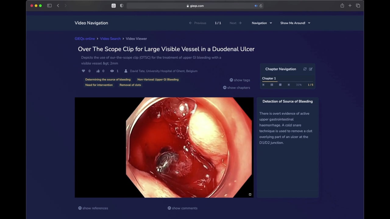
1 - Over the Scope Clip for Upper
Gastrointestinal Bleeding Use of
OTSC as first-line for life
threatening upper gastrointestinal haemorrhage.
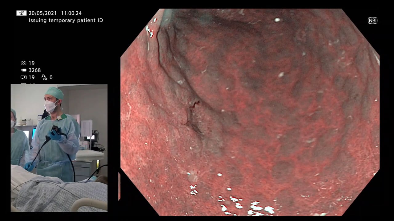
2 - Early Gastric Cancer Can you
identify and characterise
this early gastric cancer? Watch the video for more information
including endoscopic resectability
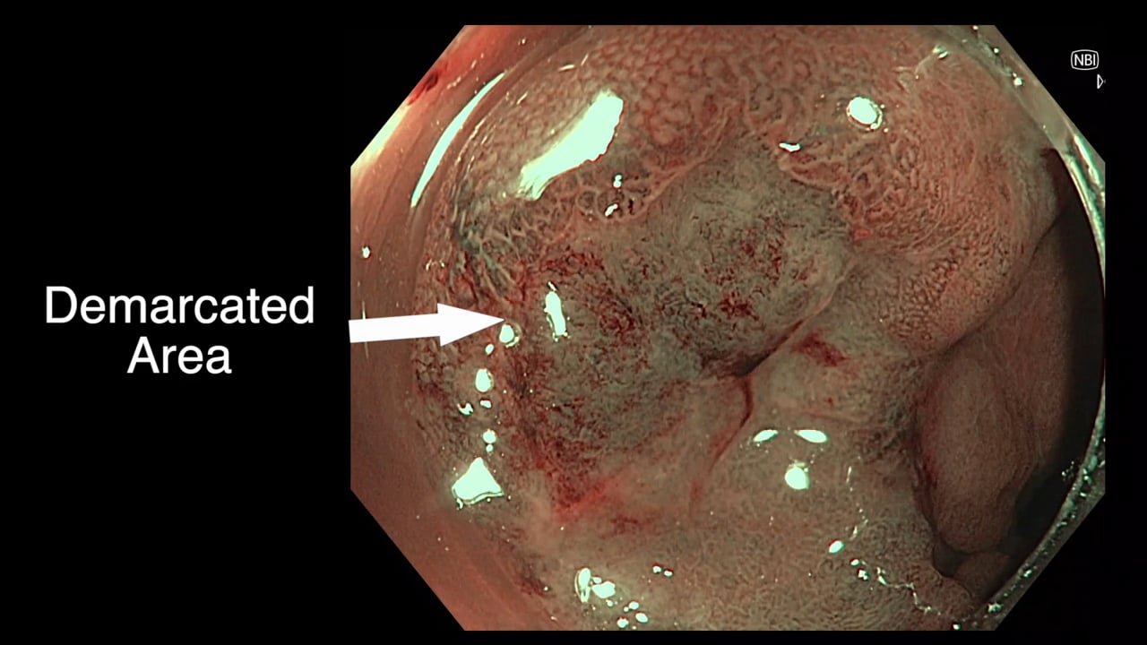
3 - The Demarcated Area as a Predictor of
Submucosal Invasion in Colon Polyps the Demarcated Area has emerged as a stable predictor
of submucosal invasive cancer. Find out more here.
4 - Dealing with Adverse Events at Colonic
Polypectomy
To be able to competently perform colonic polypectomy you must be able
to deal with adverse events. A deconstructed example is shown
here.
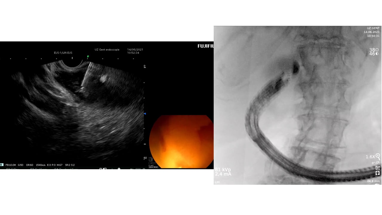
5 - Complex EUS applications to make Everyday
ERCP easier Endoscopic Ultrasound
is radically changing the way we approach biliary intervention and can
make a difference to everyday endoscopic problems.
6 - Decision Making after Large perforation and
life threatening Bleeding during Polypectomy Many of the GIEQs faculty spend their normal working
lives on complex endoscopy. Learning the lessons and approach from these
procedures, deconstructing them and bringing them to the everyday is a
crucial part of the GIEQs approach.