[{"id":"973","split":"1","chapterid":"5525","timeFrom":"0 ","timeTo":"20 ","number":"1","chaptername":"Introduction ","description":" ","tagid":"257","tagName":"Endoscopic Video and Room Video with Audio"},{"id":"973","split":"1","chapterid":"5525","timeFrom":"0 ","timeTo":"20 ","number":"1","chaptername":"Introduction ","description":" ","tagid":"1036","tagName":"Video Short"}]
[{"name":"Detecting and Managing Oesophageal Abnormalities: Key Insights and Strategies.","description":"The video discusses an oesophageal abnormality, emphasizing the importance of thorough examination, virtual chromoendoscopy imaging, biopsy, and multidisciplinary evaluation for proper management.","summary":"
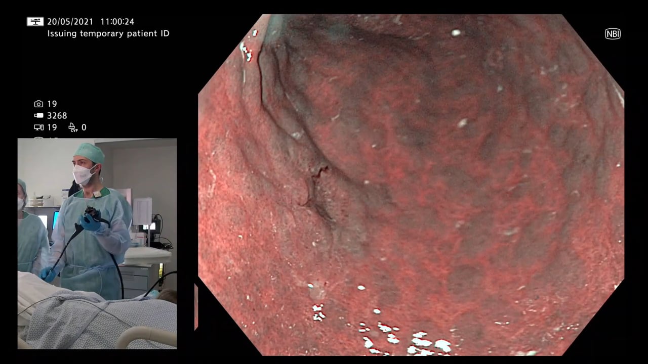
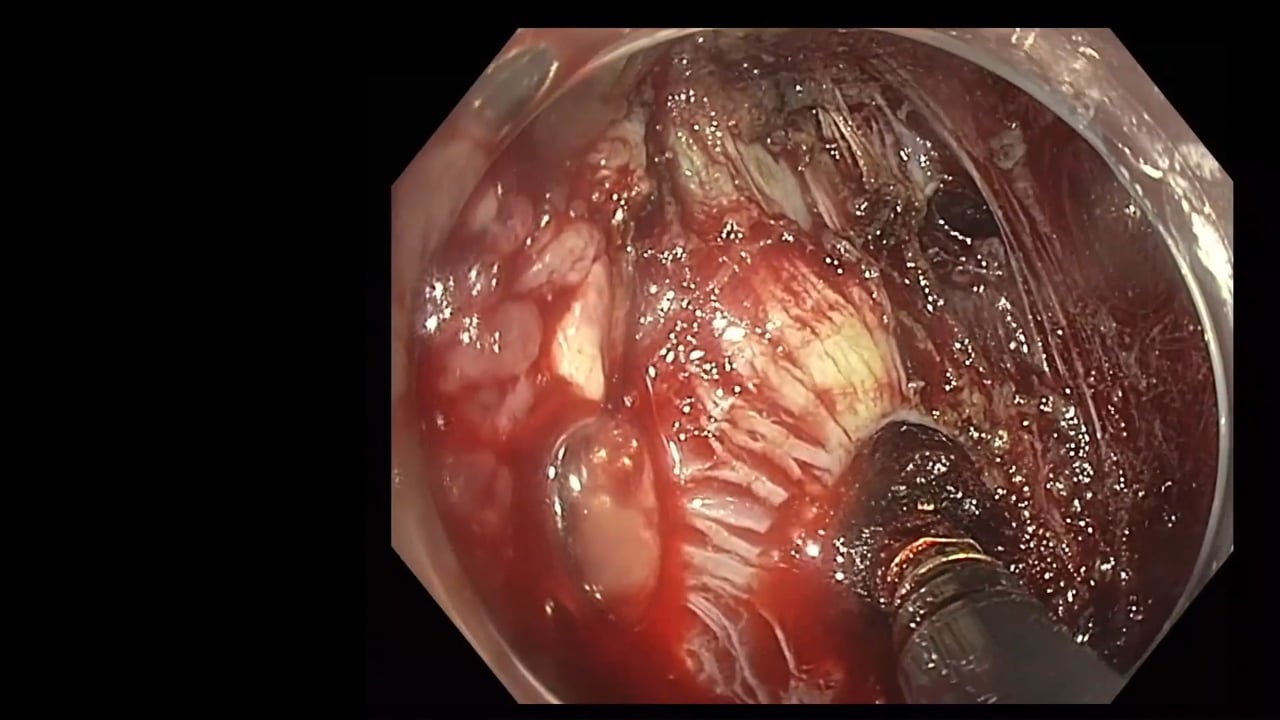
In the video, the speaker discusses an abnormality found in the oesophagus during an endoscopic examination. The abnormality appears nodular. Using virtual chromoendoscopy (NBI), the speaker emphasizes the importance of visualizing the abnormality, as it may not be easily seen in white light. The lesion is located in the mid oesophagus, extending down with difficult to define borders. It is irregular and disordered, particularly evident in the intra-papillary capillary loop (IPCL) classification using NBI. The speaker recommends capturing clear images and videos of the lesion, focusing on the most<\/span> disordered part. They stress the importance of biopsy and multidisciplinary discussion for proper evaluation and management. Thorough examination, enhanced imaging, and effective communication are crucial in these cases.<\/span> <\/span><\/p>","detailedSummary":"
· Understand the significance of thorough examination and enhanced imaging techniques in detecting oesophageal abnormalities. <\/p>\n
· Recognize the endoscopic imaging characteristics of early squamous cancer in the oesophagus. <\/p>\n
· Appreciate the value of virtual chromoendoscopy in visualizing abnormalities not easily visible in white light. <\/p>\n
· Emphasize the importance of documenting and communicating clear images and videos of the lesion, focusing on the most disordered part.<\/p>\n
· Highlight the necessity of biopsy and multidisciplinary team involvement for proper evaluation and management of such oesophageal abnormalities.<\/p>","author":"David Tate","tagger":"1","editor":"1","recorder":"1","authorid":"1","centreName":"University Hospital of Ghent","centreCity":"Ghent","centreCountry":"Belgium"}]
[{"chapterTagid":"8075","tagName":"Endoscopic Video and Room Video with Audio","id":"257"},{"chapterTagid":"8236","tagName":"Video Short","id":"1036"}]
Detecting and Managing Oesophageal Abnormalities: Key Insights and Strategies.
The video discusses an oesophageal abnormality, emphasizing the importance of thorough examination, virtual chromoendoscopy imaging, biopsy, and multidisciplinary evaluation for proper management.
In the video, the speaker discusses an abnormality found in the oesophagus during an endoscopic examination. The abnormality appears nodular. Using virtual chromoendoscopy (NBI), the speaker emphasizes the importance of visualizing the abnormality, as it may not be easily seen in white light. The lesion is located in the mid oesophagus, extending down with difficult to define borders. It is irregular and disordered, particularly evident in the intra-papillary capillary loop (IPCL) classification using NBI. The speaker recommends capturing clear images and videos of the lesion, focusing on the most disordered part. They stress the importance of biopsy and multidisciplinary discussion for proper evaluation and management. Thorough examination, enhanced imaging, and effective communication are crucial in these cases.
Detailed Summary
· Understand the significance of thorough examination and enhanced imaging techniques in detecting oesophageal abnormalities.
· Recognize the endoscopic imaging characteristics of early squamous cancer in the oesophagus.
· Appreciate the value of virtual chromoendoscopy in visualizing abnormalities not easily visible in white light.
· Emphasize the importance of documenting and communicating clear images and videos of the lesion, focusing on the most disordered part.
· Highlight the necessity of biopsy and multidisciplinary team involvement for proper evaluation and management of such oesophageal abnormalities.
Registration will open in late January 2020. Prior to this you
can register your interest below and we will keep you updated on everything GIEQs.Your email address will only be used to update you on GIEQs
Join us for GIEQs II
Released prior to the early bird deadline these 6, 1-2 minute video
snippets
demonstrate the attention to detail, deconstructed approach and rock solid evidence
base of the GIEQs Approach.
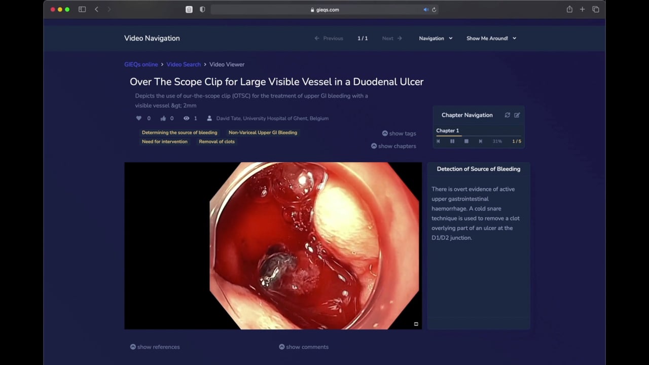
1 - Over the Scope Clip for Upper
Gastrointestinal Bleeding Use of
OTSC as first-line for life
threatening upper gastrointestinal haemorrhage.
2 - Early Gastric Cancer Can you
identify and characterise
this early gastric cancer? Watch the video for more information
including endoscopic resectability
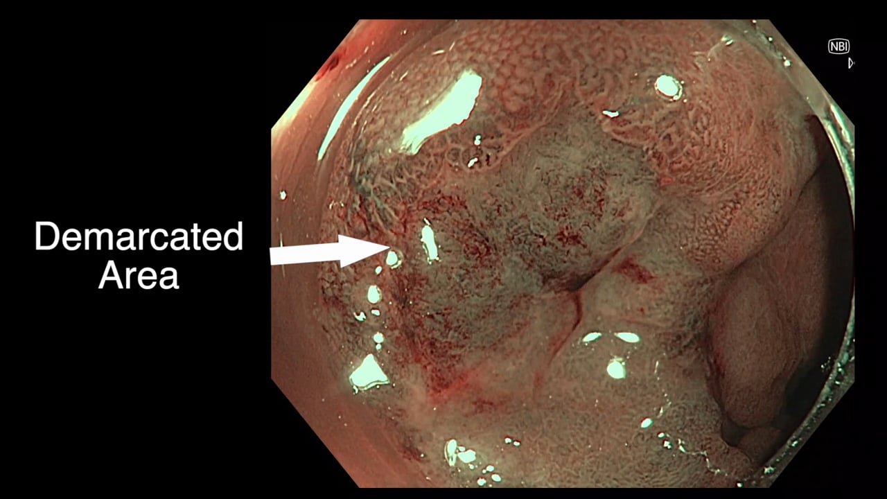
3 - The Demarcated Area as a Predictor of
Submucosal Invasion in Colon Polyps the Demarcated Area has emerged as a stable predictor
of submucosal invasive cancer. Find out more here.
4 - Dealing with Adverse Events at Colonic
Polypectomy
To be able to competently perform colonic polypectomy you must be able
to deal with adverse events. A deconstructed example is shown
here.
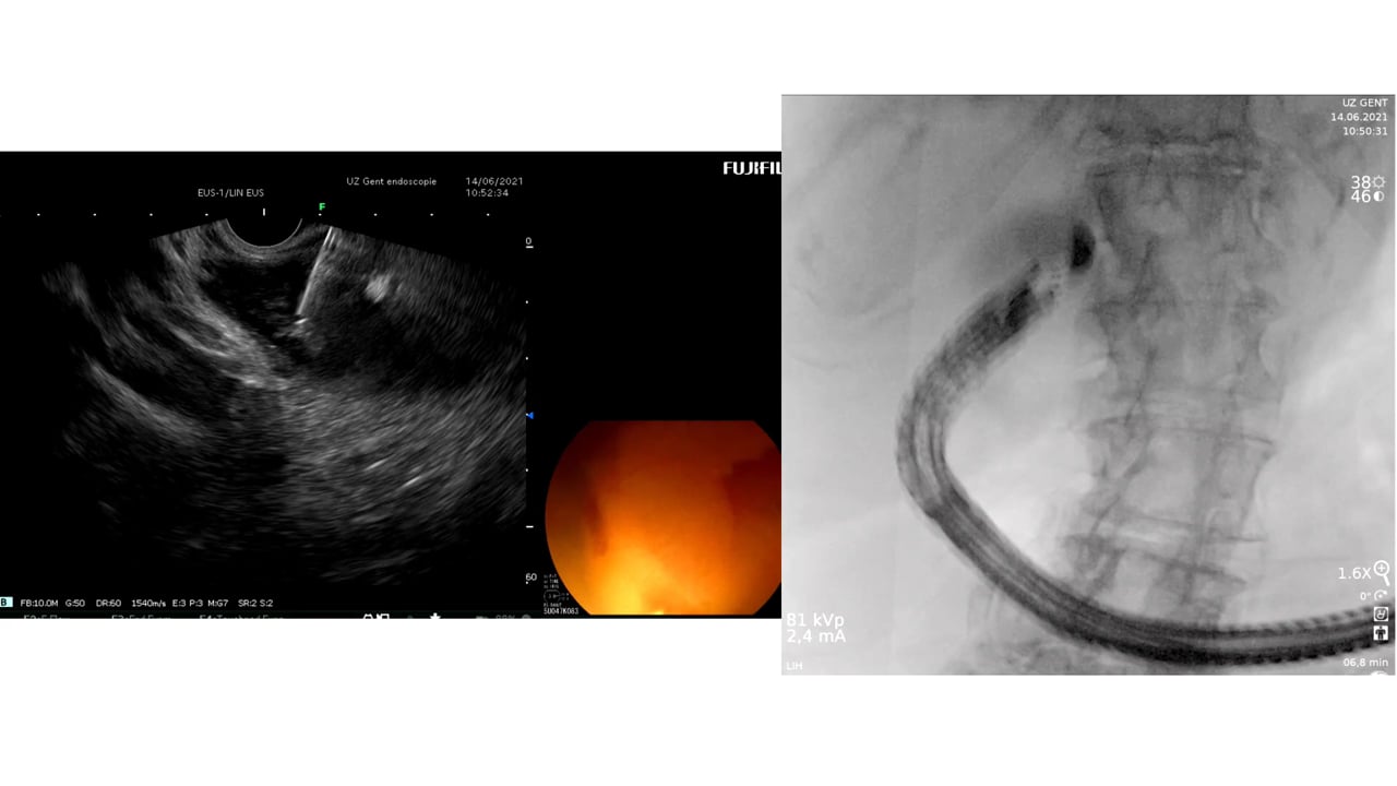
5 - Complex EUS applications to make Everyday
ERCP easier Endoscopic Ultrasound
is radically changing the way we approach biliary intervention and can
make a difference to everyday endoscopic problems.
6 - Decision Making after Large perforation and
life threatening Bleeding during Polypectomy Many of the GIEQs faculty spend their normal working
lives on complex endoscopy. Learning the lessons and approach from these
procedures, deconstructing them and bringing them to the everyday is a
crucial part of the GIEQs approach.