[{"id":"1174","chapterid":"7153","timeTo":"20.99 ","timeFrom":"6 ","number":"1","chaptername":"Esophagus Issues ","description":"In this chapter, the focus is on the chronic inflammation and stenosis observed in the oesophagus, specifically at a site that has undergone radiotherapy for a squamous cell carcinoma. The detailed exploration of the oesophagus, beginning from the upper section and moving down to the affected area at 33 centimeters, provides valuable insights into post-treatment changes such as stenosis due to the chronic inflammation following radiotherapy."},{"id":"1174","chapterid":"7154","timeTo":"54.99 ","timeFrom":"21 ","number":"2","chaptername":"Surveillance & Biopsies ","description":"In this chapter, the focus is on the management of patients entering surveillance following detection of high-grade squamous dysplasia in the oesophagus. It is demonstrated that precise navigation around the Z line is crucial for effective biopsy collection. The chapter highlights the importance of thorough visualization methods to ensure comprehensive assessment of interesting lesions and possible relative stenosis identified during the endoscopic examination."},{"id":"1174","chapterid":"7155","timeTo":"125.99 ","timeFrom":"55 ","number":"3","chaptername":"Abnormalities & Insufflation ","description":"In this chapter, a detailed evaluation of oesophageal stenosis is demonstrated, with a focus on positioning and the clockface orientation of lesions. Virtual chromoendoscopy via the Olympus 1500 platform is used to assess abnormal areas, highlighting keratinization at specific orientations, which may require further investigation. The discussion emphasizes the importance of correct positional assessment in detecting and evaluating oesophageal lesions."},{"id":"1174","chapterid":"7156","timeTo":"254.99 ","timeFrom":"126 ","number":"4","chaptername":"Resection Planning ","description":"In this chapter, the technique of endoscopic marking for resection is explored, emphasizing the strategic application of Lugol's iodine to highlight squamous dysplasia without compromising marking accuracy. The challenges of marking near stenosis are discussed, along with the use of near focus for clearer margin visualization. The approach balances traditional and virtual chromoendoscopy tools to optimize procedure outcomes."},{"id":"1174","chapterid":"7157","timeTo":"600.99 ","timeFrom":"255 ","number":"5","chaptername":"Procedure Evaluation ","description":"This chapter examines the intricate process of endoscopic evaluation, emphasizing the importance of identifying dysplastic areas and preventing stenosis through staged procedures. Specific attention is paid to visualizing abnormal IPCL patterns, utilizing imaging techniques, and assessing keratosis. Challenges in differentiating dysplasia from cancer, especially post-radiotherapy, are discussed. Techniques for marking, inflating loops, and applying contrast agents like iodine are highlighted to enhance visualization and decision-making."},{"id":"1174","chapterid":"7158","timeTo":"693 ","timeFrom":"601 ","number":"6","chaptername":"Future Considerations ","description":"This chapter discusses the examination of certain unstained areas during endoscopy, emphasizing the complexities of assessing boundaries. It highlights the role of AI in enhancing decision-making, recommending a further look at suspect areas and considering the full circumference for examination. The narrative underscores the importance of additive information techniques in improving assessment accuracy, suggesting that future advancements in AI will significantly aid endoscopists in such complex evaluations."}]
[{"id":"1174","split":"1","chapterid":"7153","timeFrom":"6 ","timeTo":"20.99 ","number":"1","chaptername":"Esophagus Issues ","description":"In this chapter, the focus is on the chronic inflammation and stenosis observed in the oesophagus, specifically at a site that has undergone radiotherapy for a squamous cell carcinoma. The detailed exploration of the oesophagus, beginning from the upper section and moving down to the affected area at 33 centimeters, provides valuable insights into post-treatment changes such as stenosis due to the chronic inflammation following radiotherapy.","tagid":"880","tagName":"Marking pre-ESD"},{"id":"1174","split":"1","chapterid":"7153","timeFrom":"6 ","timeTo":"20.99 ","number":"1","chaptername":"Esophagus Issues ","description":"In this chapter, the focus is on the chronic inflammation and stenosis observed in the oesophagus, specifically at a site that has undergone radiotherapy for a squamous cell carcinoma. The detailed exploration of the oesophagus, beginning from the upper section and moving down to the affected area at 33 centimeters, provides valuable insights into post-treatment changes such as stenosis due to the chronic inflammation following radiotherapy.","tagid":"255","tagName":"Endoscopic Video only"},{"id":"1174","split":"1","chapterid":"7154","timeFrom":"21 ","timeTo":"54.99 ","number":"2","chaptername":"Surveillance & Biopsies ","description":"In this chapter, the focus is on the management of patients entering surveillance following detection of high-grade squamous dysplasia in the oesophagus. It is demonstrated that precise navigation around the Z line is crucial for effective biopsy collection. The chapter highlights the importance of thorough visualization methods to ensure comprehensive assessment of interesting lesions and possible relative stenosis identified during the endoscopic examination.","tagid":"425","tagName":"Endoscopic Submucosal Dissection (ESD)"}]
[{"name":"Recurrent Oesophageal Squamous Dysplasia after Radiotherapy.","description":"The video explores a case of chronic esophagus inflammation with stenosis, resulting from previous radiotherapy for squamous cell carcinoma. The patient undergoes surveillance, leading to the detection of high-grade squamous dysplasia through biopsies. The video further delves into the complexities of the procedure, including marking, resection, and confirmation with Lugol's iodine. The potential role of AI in future cancer detection is also highlighted.","summary":"
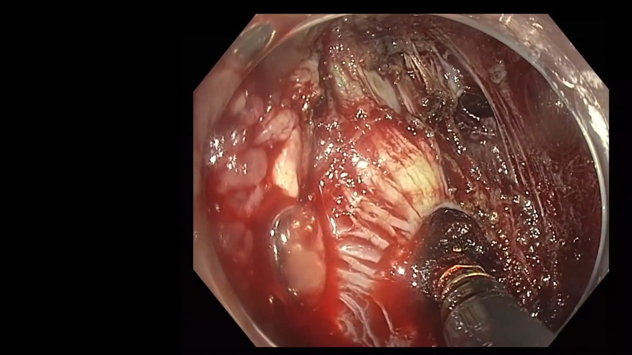
In this video, an examination of the esophagus, showing chronic inflammation and relative stenosis post-radiotherapy for squamous cell carcinoma, is demonstrated. At 33 cm from the incisors, the esophagus exhibits high-grade squamous dysplasia confirmed by biopsy. The procedure involves the use of virtual chromoendoscopy with Olympus 1500 platform and bright NBI to discern the extent of abnormality and mark the lesion for resection. Challenges in demarcating due to the extensive nature of the lesion are discussed. The application of Lugol's iodine after marking enhances the visualization of squamous dysplasia. Caution is advised during the marking to avoid inaccurate assessment which might complicate resection. Emphasis is placed on ensuring adequate insufflation and using dual-focus endoscopes for clear visualization of the margins. The importance of consideration for potential stenosis, especially in patients with a history of radiotherapy, is underscored. The role of AI in improving dysplasia detection and aiding in decision making is touched upon. Finally, particular techniques for marking are demonstrated, including the use of the tip of a knife and potential adjustments based on virtual chromoendoscopy outcomes.<\/p>","detailedSummary":"
Learning Objectives :<\/p>\n
\n
Identifying chronic inflammation and stenosis in the esophagus post-radiotherapy.<\/li>\n
Recognizing high-grade squamous dysplasia via biopsy.<\/li>\n
Utilizing virtual chromoendoscopy for enhanced visualization.<\/li>\n
Techniques for marking lesions accurately.<\/li>\n
Application of Lugol's iodine for confirming squamous dysplasia.<\/li>\n
Challenges in demarcation of extensive lesions.<\/li>\n
Importance of adequate insufflation and dual-focus scopes..<\/li>\n
Management strategies to prevent stenosis post-resection.<\/li>\n
Role of AI in enhancing detection and decision-making in endoscopy.<\/li>\n
Adjusting procedural techniques based on imaging outcomes.<\/li>\n<\/ul>","author":"David Tate","tagger":"1","editor":"1","recorder":"1","authorid":"1","centreName":"University Hospital of Ghent","centreCity":"Ghent","centreCountry":"Belgium"}]
[{"chapterTagid":"23234","tagName":"Endoscopic Submucosal Dissection (ESD)","id":"425"},{"chapterTagid":"23233","tagName":"Endoscopic Video only","id":"255"},{"chapterTagid":"23232","tagName":"Marking pre-ESD","id":"880"}]
Recurrent Oesophageal Squamous Dysplasia after Radiotherapy.
The video explores a case of chronic esophagus inflammation with stenosis, resulting from previous radiotherapy for squamous cell carcinoma. The patient undergoes surveillance, leading to the detection of high-grade squamous dysplasia through biopsies. The video further delves into the complexities of the procedure, including marking, resection, and confirmation with Lugol's iodine. The potential role of AI in future cancer detection is also highlighted.
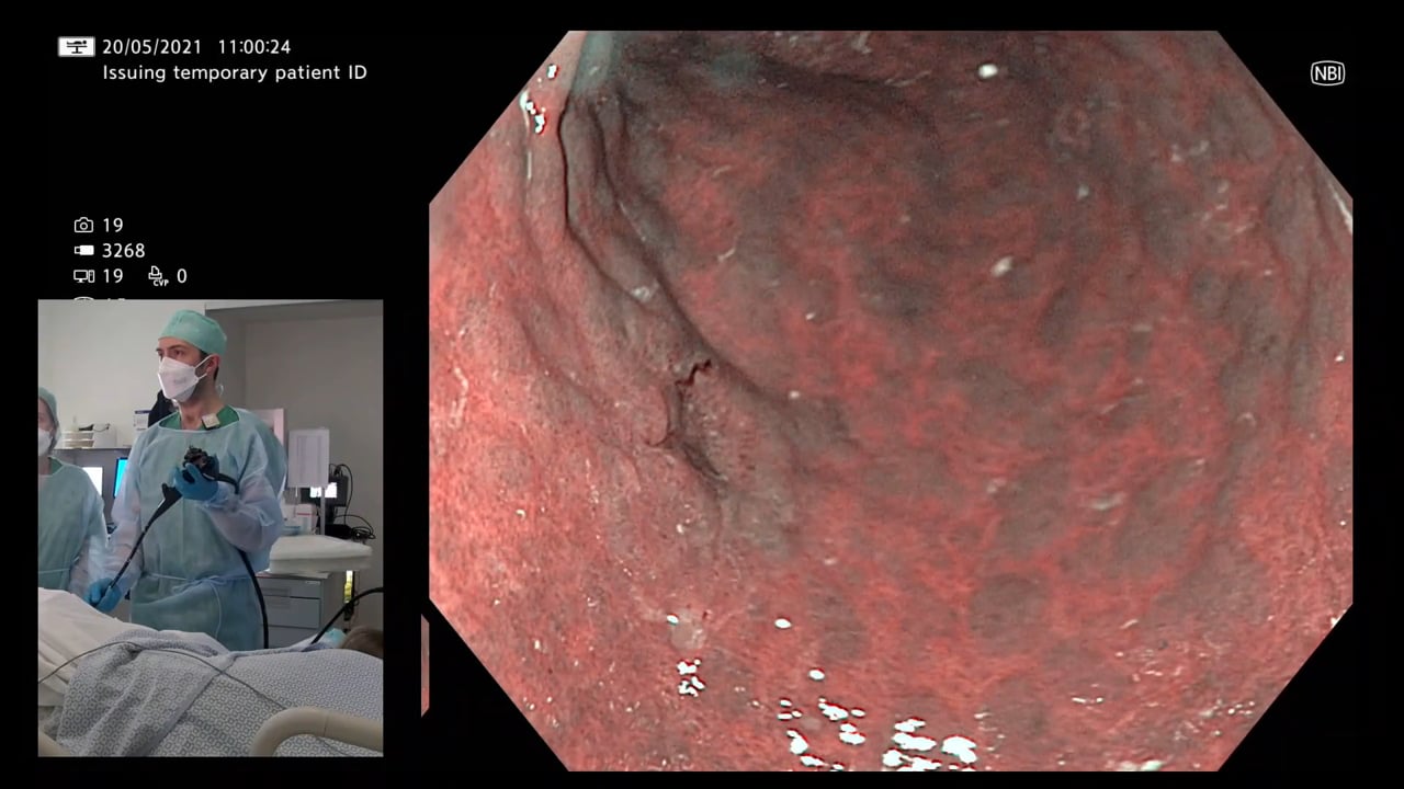
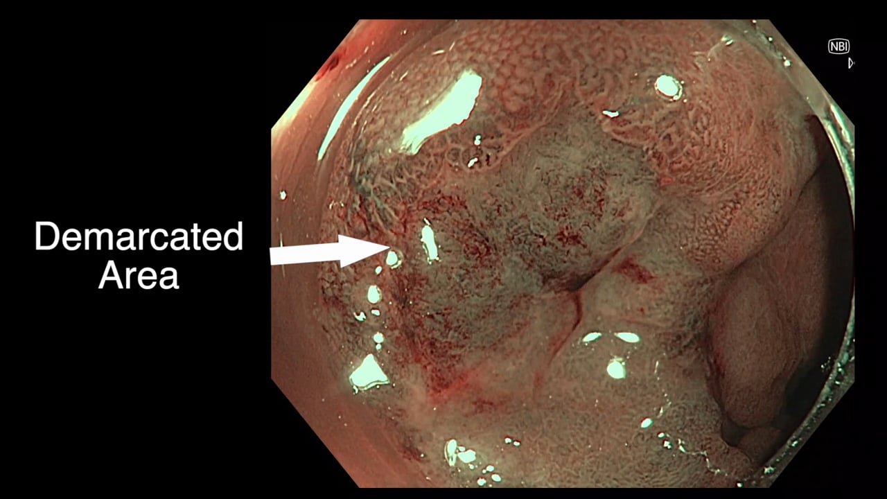
In this video, an examination of the esophagus, showing chronic inflammation and relative stenosis post-radiotherapy for squamous cell carcinoma, is demonstrated. At 33 cm from the incisors, the esophagus exhibits high-grade squamous dysplasia confirmed by biopsy. The procedure involves the use of virtual chromoendoscopy with Olympus 1500 platform and bright NBI to discern the extent of abnormality and mark the lesion for resection. Challenges in demarcating due to the extensive nature of the lesion are discussed. The application of Lugol's iodine after marking enhances the visualization of squamous dysplasia. Caution is advised during the marking to avoid inaccurate assessment which might complicate resection. Emphasis is placed on ensuring adequate insufflation and using dual-focus endoscopes for clear visualization of the margins. The importance of consideration for potential stenosis, especially in patients with a history of radiotherapy, is underscored. The role of AI in improving dysplasia detection and aiding in decision making is touched upon. Finally, particular techniques for marking are demonstrated, including the use of the tip of a knife and potential adjustments based on virtual chromoendoscopy outcomes.
Detailed Summary
Learning Objectives :
Identifying chronic inflammation and stenosis in the esophagus post-radiotherapy.
Recognizing high-grade squamous dysplasia via biopsy.
Utilizing virtual chromoendoscopy for enhanced visualization.
Techniques for marking lesions accurately.
Application of Lugol's iodine for confirming squamous dysplasia.
Challenges in demarcation of extensive lesions.
Importance of adequate insufflation and dual-focus scopes..
Management strategies to prevent stenosis post-resection.
Role of AI in enhancing detection and decision-making in endoscopy.
Adjusting procedural techniques based on imaging outcomes.
Registration will open in late January 2020. Prior to this you
can register your interest below and we will keep you updated on everything GIEQs.Your email address will only be used to update you on GIEQs
Join us for GIEQs II
Released prior to the early bird deadline these 6, 1-2 minute video
snippets
demonstrate the attention to detail, deconstructed approach and rock solid evidence
base of the GIEQs Approach.
1 - Over the Scope Clip for Upper
Gastrointestinal Bleeding Use of
OTSC as first-line for life
threatening upper gastrointestinal haemorrhage.
2 - Early Gastric Cancer Can you
identify and characterise
this early gastric cancer? Watch the video for more information
including endoscopic resectability
3 - The Demarcated Area as a Predictor of
Submucosal Invasion in Colon Polyps the Demarcated Area has emerged as a stable predictor
of submucosal invasive cancer. Find out more here.
4 - Dealing with Adverse Events at Colonic
Polypectomy
To be able to competently perform colonic polypectomy you must be able
to deal with adverse events. A deconstructed example is shown
here.
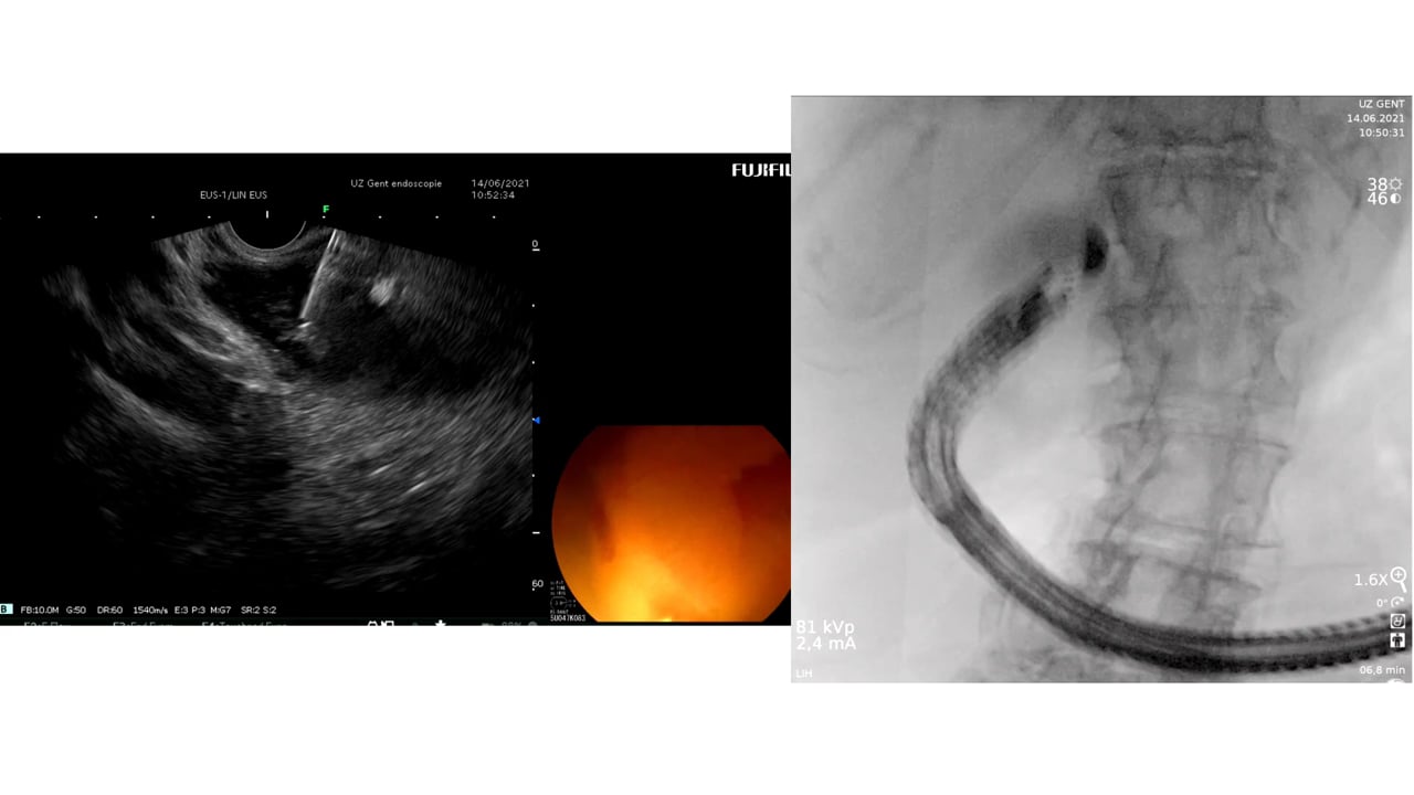
5 - Complex EUS applications to make Everyday
ERCP easier Endoscopic Ultrasound
is radically changing the way we approach biliary intervention and can
make a difference to everyday endoscopic problems.
6 - Decision Making after Large perforation and
life threatening Bleeding during Polypectomy Many of the GIEQs faculty spend their normal working
lives on complex endoscopy. Learning the lessons and approach from these
procedures, deconstructing them and bringing them to the everyday is a
crucial part of the GIEQs approach.