[{"name":"How to image Barrett's Oesophagus endoscopically","description":"Doctor's meticulous analysis of an esophageal endoscopy revealed high-grade dysplasia and Barrett's esophagus, emphasizing precise imaging techniques and tumor demarcation.","summary":"
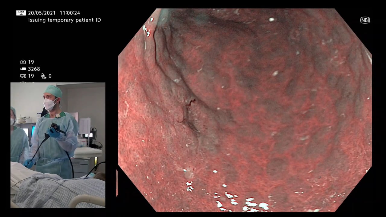
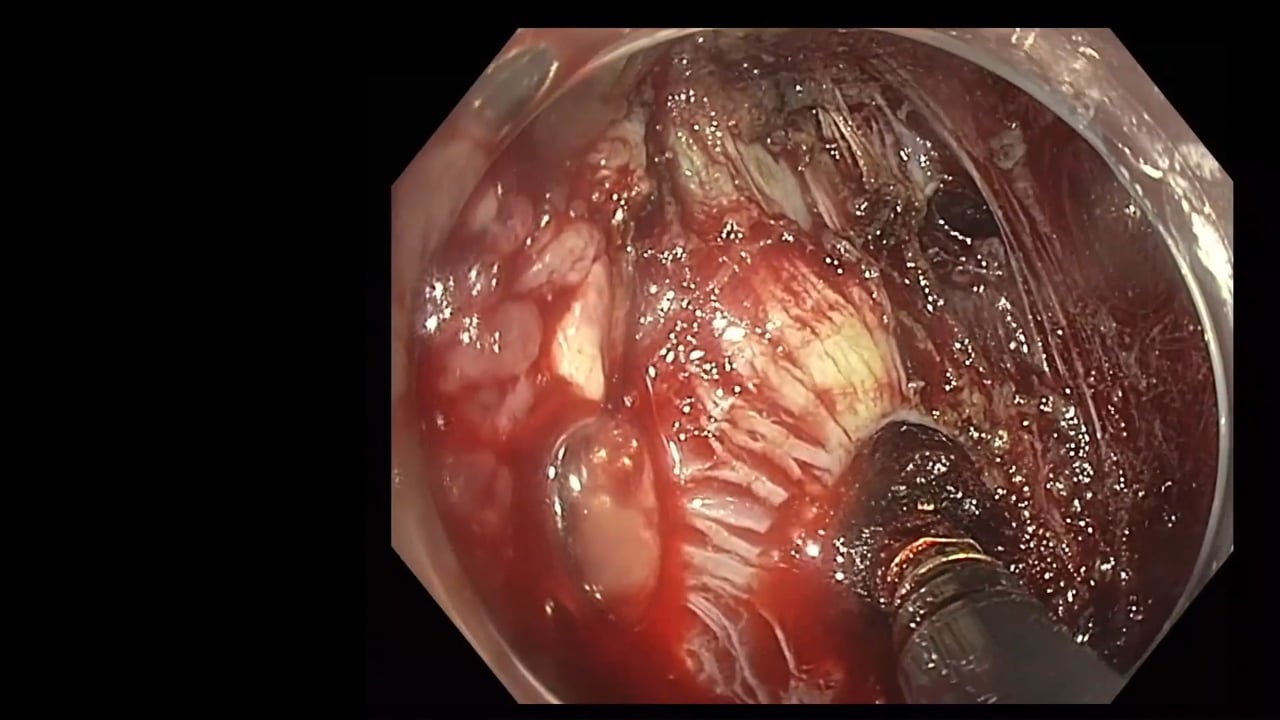
In the detailed discussion, a doctor based in Belgium expertly analyzed an endoscopic examination of the esophagus, specifically focusing on the identification of high-grade dysplasia and Barrett's esophagus. The doctor's comprehensive description showcased their clinical acumen, beginning with the seemingly normal appearance of the esophagus and gradually revealing irregularities. They emphasized the importance of virtual chromoendoscopy and precise imaging techniques to discern subtle tissue patterns and abnormalities. <\/p>\n
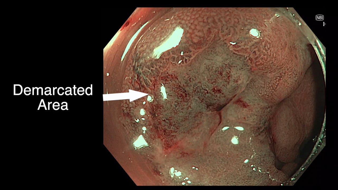
The highlight of the discussion was the clear demarcation line distinguishing the tumor from surrounding tissue, which is crucial for treatment decisions. The doctor's expertise and detailed presentation not only serve as an educational resource but also underscore their proficiency in gastroenterology and endoscopy. Overall, this insightful analysis demonstrates the doctor's valuable contributions to patient care and medical knowledge.<\/p>","detailedSummary":"
Learning Objectives:<\/p>\n
\n
Understand the progression of abnormalities in esophageal endoscopy, from apparent normality to identification of irregularities. <\/li>\n
Learn the significance of virtual chromoendoscopy in enhancing mucosal pattern visualization. <\/li>\n
Recognize the importance of controlling focal length using a cap for clearer images. <\/li>\n
Identify the demarcation line to guide treatment decisions in cases of high-grade dysplasia. <\/li>\n
Appreciate the value of detailed endoscopic analysis in gastroenterology practice. <\/li>\n<\/ul>","author":"David Tate","tagger":"1","editor":"1","recorder":"1","authorid":"1","centreName":"University Hospital of Ghent","centreCity":"Ghent","centreCountry":"Belgium"}]
Doctor's meticulous analysis of an esophageal endoscopy revealed high-grade dysplasia and Barrett's esophagus, emphasizing precise imaging techniques and tumor demarcation.
In the detailed discussion, a doctor based in Belgium expertly analyzed an endoscopic examination of the esophagus, specifically focusing on the identification of high-grade dysplasia and Barrett's esophagus. The doctor's comprehensive description showcased their clinical acumen, beginning with the seemingly normal appearance of the esophagus and gradually revealing irregularities. They emphasized the importance of virtual chromoendoscopy and precise imaging techniques to discern subtle tissue patterns and abnormalities.
The highlight of the discussion was the clear demarcation line distinguishing the tumor from surrounding tissue, which is crucial for treatment decisions. The doctor's expertise and detailed presentation not only serve as an educational resource but also underscore their proficiency in gastroenterology and endoscopy. Overall, this insightful analysis demonstrates the doctor's valuable contributions to patient care and medical knowledge.
Detailed Summary
Learning Objectives:
Understand the progression of abnormalities in esophageal endoscopy, from apparent normality to identification of irregularities.
Learn the significance of virtual chromoendoscopy in enhancing mucosal pattern visualization.
Recognize the importance of controlling focal length using a cap for clearer images.
Identify the demarcation line to guide treatment decisions in cases of high-grade dysplasia.
Appreciate the value of detailed endoscopic analysis in gastroenterology practice.
Registration will open in late January 2020. Prior to this you
can register your interest below and we will keep you updated on everything GIEQs.Your email address will only be used to update you on GIEQs
Join us for GIEQs II
Released prior to the early bird deadline these 6, 1-2 minute video
snippets
demonstrate the attention to detail, deconstructed approach and rock solid evidence
base of the GIEQs Approach.
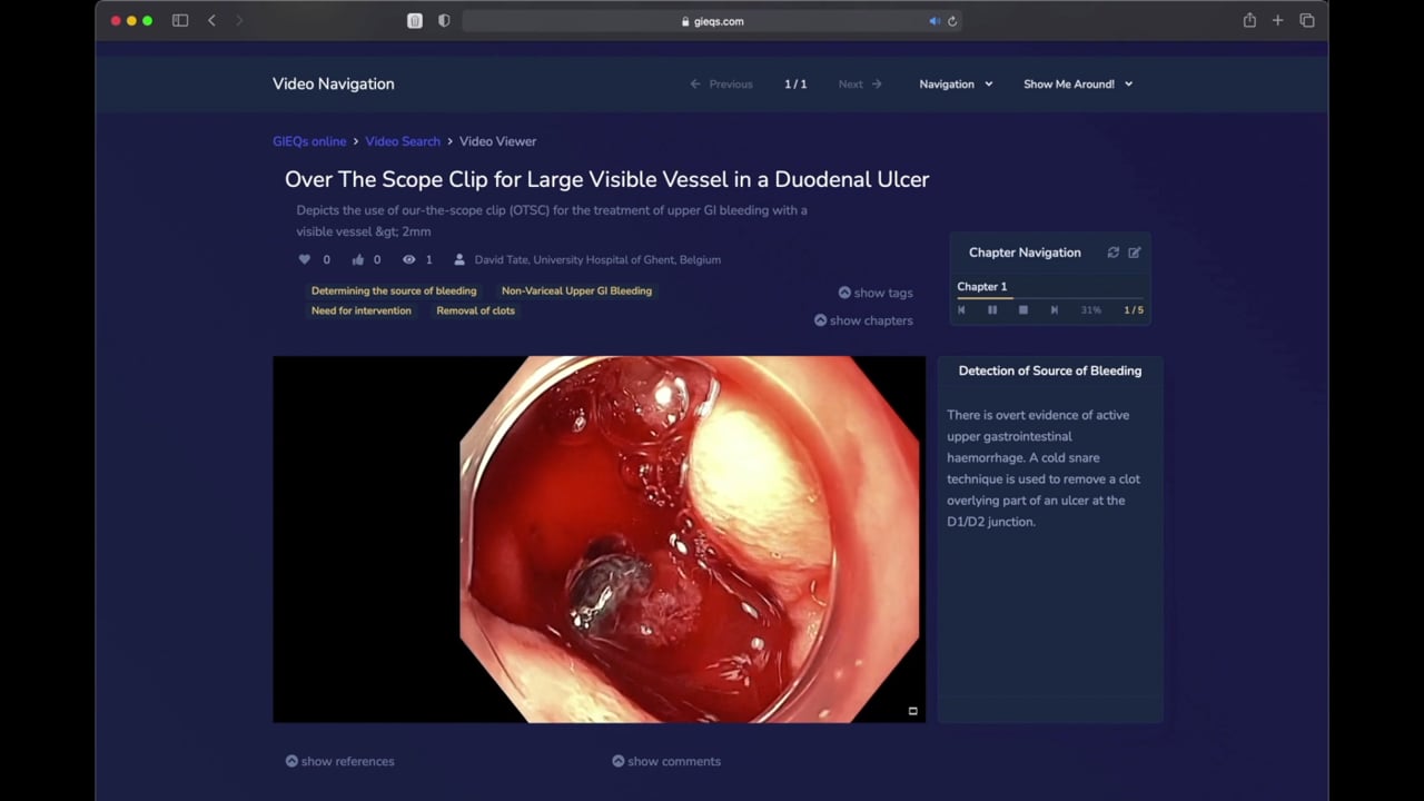
1 - Over the Scope Clip for Upper
Gastrointestinal Bleeding Use of
OTSC as first-line for life
threatening upper gastrointestinal haemorrhage.
2 - Early Gastric Cancer Can you
identify and characterise
this early gastric cancer? Watch the video for more information
including endoscopic resectability
3 - The Demarcated Area as a Predictor of
Submucosal Invasion in Colon Polyps the Demarcated Area has emerged as a stable predictor
of submucosal invasive cancer. Find out more here.
4 - Dealing with Adverse Events at Colonic
Polypectomy
To be able to competently perform colonic polypectomy you must be able
to deal with adverse events. A deconstructed example is shown
here.
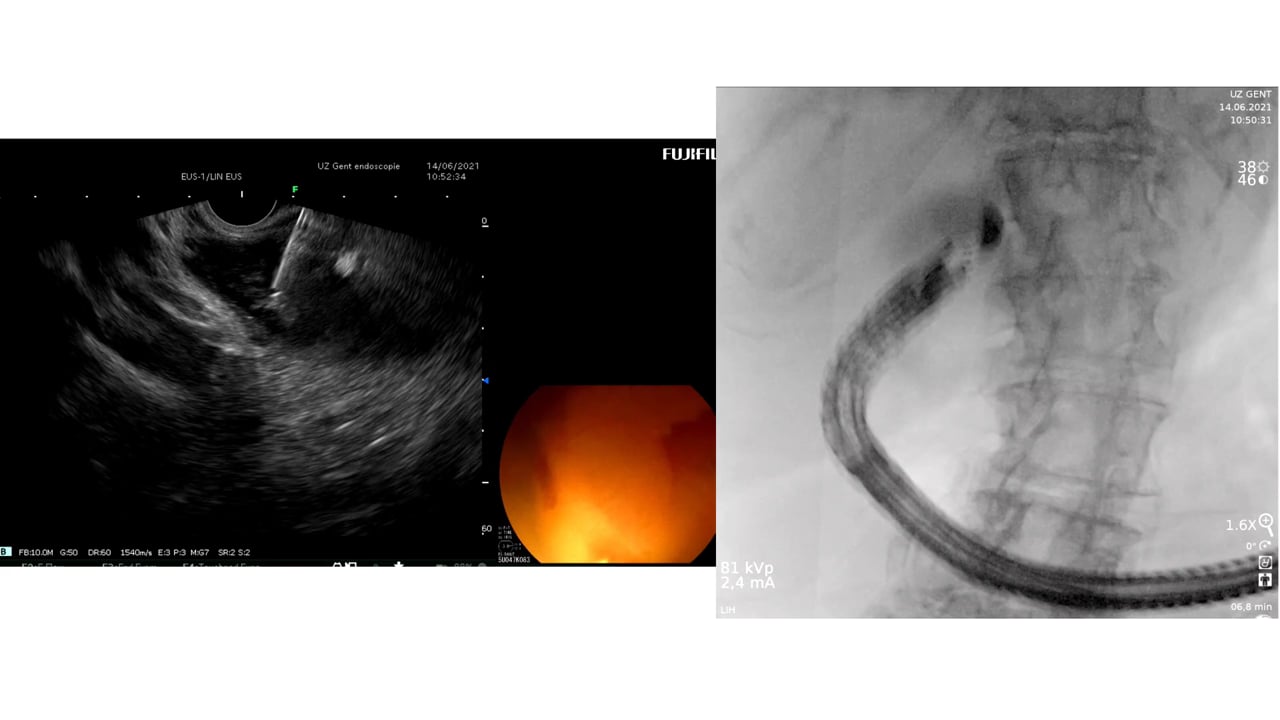
5 - Complex EUS applications to make Everyday
ERCP easier Endoscopic Ultrasound
is radically changing the way we approach biliary intervention and can
make a difference to everyday endoscopic problems.
6 - Decision Making after Large perforation and
life threatening Bleeding during Polypectomy Many of the GIEQs faculty spend their normal working
lives on complex endoscopy. Learning the lessons and approach from these
procedures, deconstructing them and bringing them to the everyday is a
crucial part of the GIEQs approach.